What is a 3D Ultrasound?
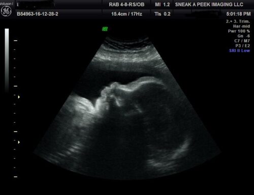
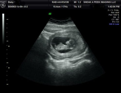
During your 3D ultrasound session we use a screening device to view the fetus during pregnancy. 3D ultrasounds are just still pictures of the fetus in three dimensions. They use high-frequency sound waves that are transmitted using a handheld probe to emit and even capture impulses produced by ultrasound waves that are emitted at different angles to come up with high resolution and clear 3D images of the baby. With the images, you can have a clear view of the umbilical cord and the placenta to access to health of the mother and baby.
How do 3D ultrasounds work?
3D ultrasound in early stages of pregnancy is performed by placing a transducer or a probe within the vagina or on the abdominal area where sounds waves are emitted at millions of cycles at a given second. For a bigger fetus, a transducer is moved around the mother’s belly at different angles to reveal the contours of baby features. The reflected echoes are then sent back to the computer for programming. The results are amazingly clear 3D volume images of the fetus surface and internal organs.
How soon can you have 3D ultrasounds?
The earliest you should have a 3D ultrasound is 25 weeks of your pregnancy. The best time for optimal images is 27-30 weeks of pregnancy.
However, in most cases the 25th week is not the ideal time for 3D ultrasounds. You should arrange your appointment between the 27th and 30th week. Reason being, at this period, the baby is nice in size, usually in a great profile position, and theres enough amniotic fluid in Moms belly! After 34 weeks the baby is likely to have descended into the pelvis which may make it harder to achieve a great level of detail that Mom would desire.
Do you need a 3D ultrasound?
No, getting a 3D ultrasound is exciting and it is gives the Mommy an opportunity to see her baby’s face and how much baby has grown! Your doctor may use 3D ultrasound to help detect any developmental abnormalities. Also, your doctor could decide to use 3D 4D ultrasound to help measure the amount of amniotic fluid, just in case the mother is losing it or not replenishing it at the normal rates. If the placenta has problems such as abruption placentae (placenta growing prematurely) and placenta previa (wrongly positioned), a 3D ultrasound may help detect it.
Other benefits of having a 3D ultrasound performed include:
– To confirm the lying position of the unborn baby
– Detect abnormalities of the uterus
– An incredible bonding experience for mother and family to be
Is 3D ultrasound safe?
All medical procedures are believed to have a risk. However, there’s no evidence to show a 3D ultrasound done in the right way will harm the mother and the unborn baby. Done properly by a trained technician, it has no known side effects.


